Compact Bone Diagram Labeled : Bone Structure Anatomy And Physiology I. 90 introduction to the skeletal system exercise 7 Compact bone is the denser, stronger of the two types of bone tissue (figure 6.12). Online quiz to learn compact bone diagram; Compact bone accounts for 80% of the bones in the human body. This video describes the microscopic anatomy of compact bone.
Compact bone accounts for 80% of the bones in the human body. Labeling portions of a long bone learn with flashcards, games and more — for free. Production of compact bone c. Good, here in this part, i am going to describe the structure of compact bone. It can be found under the periosteum and in the diaphyses of long bones, where it provides support and protection.
Reviewing Facts Terms Labeling from wps.pearsoned.com Compact bone histology slide structure with diagram. This short article will describe the spongy bone histology and labeled diagram and real slide pictures. You need to get 100% to score the 15 points available. Compact bone, also called cortical bone, dense bone in which the bony matrix is solidly filled with organic ground substance and inorganic salts, leaving only tiny spaces (lacunae) that contain the osteocytes, or bone cells.compact bone makes up 80 percent of the human skeleton; Online quiz to learn compact bone diagram; Bone marrow diagram, compact bone diagram quiz, compact bone slide labeled, diagram long bone, labeled compact bone model, human anatomy, bone marrow diagram, compact bone diagram quiz, compact bone slide labeled, diagram long bone, labeled compact bone model. There are small canals that run through the bone, which allow blood vessels to penetrate it. (b) in this micrograph of the osteon, you can clearly see the concentric lamellae and central canals.
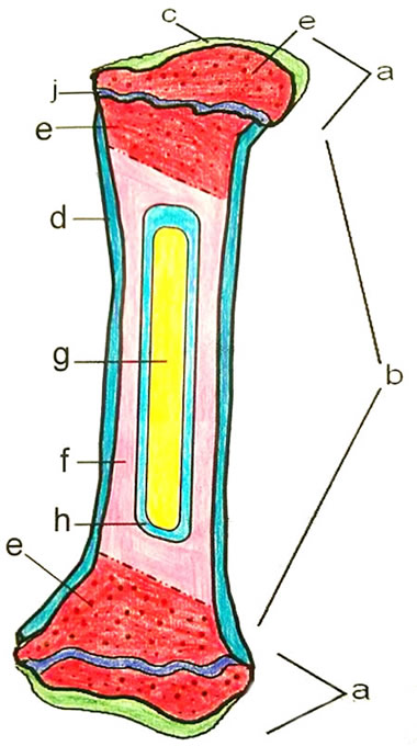
Bone cross section diagram labeled :
Stages in long bone development tissue / organ: The shafts found in long bones are also compact bones. The compact bone is composed of calcified extracellular material the bone matrix and 3 major cell types which are osteoblast which ssynthesize and secrete the organic components of bone matrix which include type 1 collagen fibers proteoglycans and several glycoproteins such as ostepnectin. What is the structure and function of the compact bone.as shown in figure 2. Good, here in this part, i am going to describe the structure of compact bone. Some, mostly older, compact bone is remodelled to form these haversian systems (or osteons). 90 introduction to the skeletal system exercise 7 Start studying compact bone labeling. The remainder is cancellous bone, which has a spongelike appearance with numerous large spaces and is found in the. This video describes the microscopic anatomy of compact bone. In long bones, as you move from the outer cortical compact bone to the inner medullary cavity, the bone transitions to spongy bone. It can be found under the periosteum and in the diaphyses of long bones, where it provides support and protection. This short article will describe the spongy bone histology and labeled diagram and real slide pictures.
There are small canals that run through the bone, which allow blood vessels to penetrate it. Terms in this set (8) spongy bone (contains red marrow) compact bone (has osteons) osteon. (b) in this micrograph of the osteon, you can clearly see the concentric lamellae and central canals. This video describes the microscopic anatomy of compact bone. Bone cross section diagram labeled :
Bone Coloring Answer Key And Coloring Sample from www.biologycorner.com The main type of bone cell is the osteocyte (bone cell, shown as purple in the diagram). Compact bone is the denser, stronger of the two types of osseous tissue (figure 6.3.6). Bone model label the parts of a compact bone. (b) in this micrograph of the osteon, you can clearly see the concentric lamellae and central canals. Start studying compact bone labeling. The cells of compact bone, which is also called cortical bone, appear to be tightly packed into a solid mass. This video describes the microscopic anatomy of compact bone. The diagram above shows a longitudinal view of an osteon.
Some, mostly older, compact bone is remodelled to form these haversian systems (or osteons).
Compact bone is the denser, stronger of the two types of osseous tissue (figure 6.3.6). You need to get 100% to score the 15 points available. The shaft is composed of compact bone this page is about compact bone labeled diagram,contains anatomy & physiology i bis 240: 12 photos of the diagram of a long bone anatomy bone function, describe the structure of a bone, diagram compact bone, diagram femur, diagram osteon, structure of bones, what does spongy bone do, human anatomy, bone function, describe the structure of a bone. Some, mostly older, compact bone is remodelled to form these haversian systems (or osteons). The endosteum can be seen in the t.s. 0 0000 a shoutout is a way of letting people know of a. The cells of compact bone, which is also called cortical bone, appear to be tightly packed into a solid mass. The shafts found in long bones are also compact bones. It can be found under the periosteum and in the diaphyses of long bones, where it provides support and protection. However, compact bones also serve a function in storing and releasing calcium to the. Long bone diagram labeled colored. Label the structural components of bone tissue in the diagram:
It can be found under the periosteum and in the diaphyses of long bones, where it provides support and protection. The compact bone is composed of calcified extracellular material the bone matrix and 3 major cell types which are osteoblast which ssynthesize and secrete the organic components of bone matrix which include type 1 collagen fibers proteoglycans and several glycoproteins such as ostepnectin. 12 photos of the diagram of a long bone anatomy bone function, describe the structure of a bone, diagram compact bone, diagram femur, diagram osteon, structure of bones, what does spongy bone do, human anatomy, bone function, describe the structure of a bone. Labeling portions of a long bone. It can be found under the periosteum and in the diaphyses of long bones, where it provides support and protection.
6 3 Bone Structure Anatomy Physiology from open.oregonstate.education In the last article, i described the compact bone histology with labeled diagram and real slide pictures. Compact bone histology slide structure with diagram. 0 0000 a shoutout is a way of letting people know of a. Start studying compact bone labeling. The endosteum can be seen in the t.s. Compact bone accounts for 80% of the bones in the human body. You need to get 100% to score the 15 points available. Compact bone is the denser, stronger of the two types of bone tissue (figure 6.12).
Long bone diagram labeled colored.
However, compact bones also serve a function in storing and releasing calcium to the. (b) in this micrograph of the osteon, you can clearly see the concentric lamellae and central canals. What is the structure and function of the compact bone.as shown in figure 2. The remainder is cancellous bone, which has a spongelike appearance with numerous large spaces and is found in the. This video describes the microscopic anatomy of compact bone. In long bones, as you move from the outer cortical compact bone to the inner medullary cavity, the bone transitions to spongy bone. Good, here in this part, i am going to describe the structure of compact bone. Some, mostly older, compact bone is remodelled to form these haversian systems (or osteons). Start studying compact bone labeling. 90 introduction to the skeletal system exercise 7 Learn vocabulary, terms, and more with flashcards, games, and other study tools. Compact bone is the denser, stronger of the two types of osseous tissue (figure 6.3.6). The compact bone is the main structure in the body for support, protection, and movement.
Due to the strong nature of compact bone, compared to spongy bone, it is the preferred tissue for strength compact bone diagram. Stages in long bone development tissue / organ:
Share :
Post a Comment
for "Compact Bone Diagram Labeled : Bone Structure Anatomy And Physiology I"


Post a Comment for "Compact Bone Diagram Labeled : Bone Structure Anatomy And Physiology I"